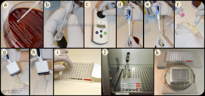
Dilutions methodsBacteria (a) are mixed with MilliQ water/NaCl (b) suspension to a turbidity of 0.5 McFarland (c). A part of the suspension (d) are mixed (e) with Mueller-Hinton Broth (f) and inoculated (g) into the wells of the microtitre plate (h), which are covered with plastic film (i) and incubated (j) before determination of MIC value (k). Image: Homayoon Davam, Alva Gustafsson, Ingrid Hanssson (HBIO, SLU). - Click on the image to enlarge it. In the dilution method, the minimum inhibitory concentration (MIC) of an antibiotic for a bacterium is determined. The method is based on the growth of bacteria on agar or broth with different concentrations of antibiotics in two-fold series in tubes or wells. For practical reasons, commercially available microtiter plates are mainly used in routine diagnostics laboratories where dilution series of different antibiotics are prepared and mostly dehydrated into a microtiter plate with 96 wells. For growth control, two of the wells are usually without antibiotics and used for positive control. Bacteria whose sensitivity against different antibiotics is to be tested are taken from a pure culture and homogenised with a supsension of ultra clean water or NaCl to obtain a fixed concentration of bacterial suspension, which means a turbidity of 0.5 McFarland. A part of the suspension is then mixed with Mueller-Hinton broth to obtain the desired concentration (104-105 bacteria/ml). For weak and slow growing bacteria, 5% serum is mostly added to achieve the desired growth. Thereafter a standard volume of this bacterial suspension is inoculated in each well and the microtiter plate is sealed with plastic and incubated for the optimal culture conditions of the tested bacteria, such as time, temperature and atmosphere. After incubation, the growth control is visually checked to ensure growth in the wells that do not contain antibiotics. Thereafter, growth or not growth is read in the different wells with antibiotics. In the well with the lowest concentration of antimicrobials that completely inhibits bacterial growth detected by the eye is decided as the MIC expressed in mg/L or μg/ml. To determine the sensitivity of the bacterium, the MIC value obtained is compared with breakpoints (ECOFF) for current antibiotics in of the scientific committees´guidelines for antimicrobial resistance, such as the European Committee on Antimicrobial Susceptibility Testing (EUCAST, https://www.eucast.org/mic_distridutions_and ecoffs). Another dilution method used for testing susceptibility to antibiotics is the Epsilometer-test (E-test) where the MIC is determined by using gradient strips impregnated with predefined exponential gradient of antibiotic. The bacteria whose susceptibility is to be tested is inoculated as a lawn on an agar plate, usually Mueller Hinton agar with or without blood. After inoculation of the bacterial suspension, the strip with the antimicrobial concentration gradient are immediately places on the inoculated agar and incubated for 18-24 hours. The E-test strip begin immediately to release antibiotics when applied on the inoculated agar plate. After incubation, a symmetrical inhibition ellipse centred along the strip is visible if the bacteria is sensitive to the antibiotic. The MIC is determined where the zone of the inhibition intersect the interpretive scale printed on the strip. If the bacteria is resistant to the tested antimicrobial substance the growth occurs along the entire strip. Updated: 2025-05-13. |

